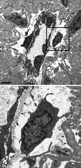
Fig. 1.

Electron micrograph showing the morphological arrangement of a large P10 RON capillary (box shown at higher magnification below). The capillary lumen is 2–8 μm in diameter and is filled with plasma which appears grainy. The vessel is surrounded by astrocyte processes which cover the outer basement membrane, by axon profiles which are largely pre-myelinated and by oligodendroglial (‘Ol’) and astrocyte (‘As’) somata. Three endothelial somata (‘En’) and their processes line the lumen and are surrounded by an inner basement membrane (arrows). Two pericyte somata and their processes are located between the inner and outer (arrow heads) basement membrane. Pericyte inclusions include a wide-bore endoplasmic reticulum (‘*’), mitochondria and cytoplasmic granules. Note the fine processes extending from the larger pericyte processes. Scale bar: 2 μm.
