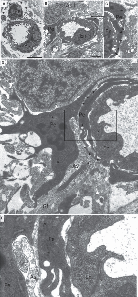
Fig. 2.

Electron micrographs showing the typical morphological arrangement of small P10 RON capillaries. (A) A P10 RON capillary with a lumen diameter of 2–3 μm. The somata and processes of a single endothelial cell (‘En’) line the capillary lumen and the somata and processes of a pericyte (‘Pe’) are located under the basement membrane (arrowheads). (B,C) A P10 capillary with a luminal diameter of 3–4 μm (boxed area in B is shown in C). An endothelial cell is enclosed within a pericyte process under the basement membrane (C, arrowheads). The pericyte process makes close connections with the endothelia cell (‘C’, arrows). (D,E) Same section as B but at higher magnification. The pericyte somata is next to the capillary basement membrane and extends processes to nearby glial cells and their processes. A perivascular process merges with the basement membrane (D, arrowheads), while an astrocyte process (As) containing glial filaments (Gf) intervenes between the pericyte somata and the basement membrane. The boxed area in D is shown at higher gain in ‘E’, revealing that the pericyte contains wide-bore, rough, branched endoplasmic reticulum, while the endothelial cell contains narrow-bore, smooth, endoplasmic reticulum (*). The endothelia cell also contains numerous cytoplasmic granules and vesicular bodies. Scale bar: 2 μm.
