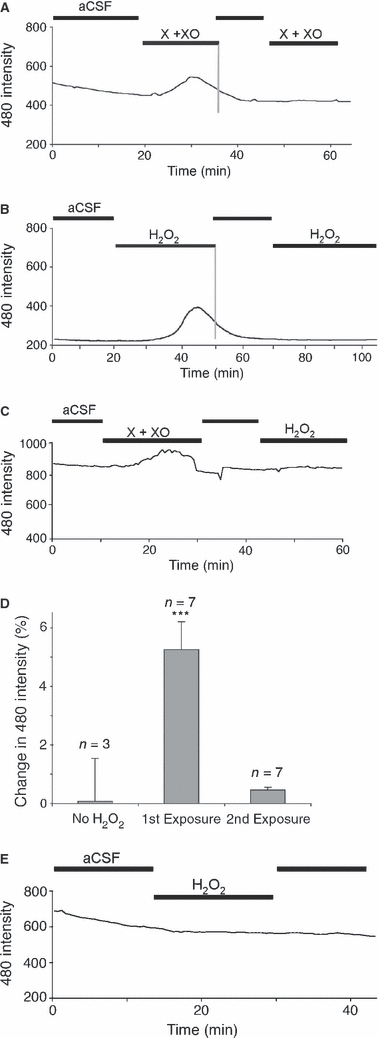
Fig. 4.

Exogenous ROS evoke transient intracellular ROS rises in neonatal rat optic nerve. (A) Intracellular ROS levels are assessed as changes in DCF fluorescence (following excitation at 480 nm) in H2-DCDFD-loaded P10 optic nerve. There is a gradual decline in fluorescence with time in aCSF and an elevation following the application of xanthine + xanthine oxidase (X + XO). The fluorescent signal falls back to baseline in the continued presence of X + XO and a second application has no effect. (B) A similar series of events are seen in this optic nerve following the application of 100 μm H2O2. (C) An exposure to X + XO blocks any subsequent effect of H2O2 exposure. (D) Amplitude of DCF fluorescence changes in control nerves, following a first exposure to 100 μm H2O2 or following a second exposure (protocol as in Fig. 2B). ***P < 0.001 vs. control. (E) An optic nerve exposed to 100 μm H2O2 in a Petri dish for 20 min prior to washing; H2-DCDFD loading shows no response to a subsequent 100 μm H2O2 exposure. All data plots show single representative experiments.
