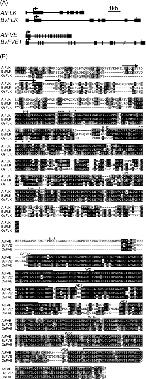
Fig. 1.
Sequence and structure of the autonomous pathway gene homologues BvFLK and BvFVE1. (A) Exon–intron structure of BvFLK, BvFVE1, and the respective A. thaliana genes (FLK, accession number AAX51268; FVE, accession number AF498101). Exons are indicated as black rectangles, and the position of start and stop codons is indicated by arrows and vertical bars, respectively. (B) Pairwise sequence alignments and domain organization. The alignments were generated using ClustalW2 (http://www.ebi.ac.uk/Tools/clustalw2/index.html). Identical and similar residues are highlighted by black or grey boxes, respectively. The position of protein domains according to Pfam 22.0 (http://pfam.sanger.ac.uk/) is marked by horizontal lines above the alignment. In FLK, the positions of three perfect eight-residue repeats and the core residues of K-homology RNA-binding (KH) domains (Mockler et al., 2004) are indicated by arrows and asterisks, respectively. The first and sixth WD40 repeat domains (WD1 and WD6) were not identified by Pfam and were annotated according to Ausin et al. (2004). A putative nuclear localization signal (NLS; black line) in FVE according to Ausin et al. (2004) and a potential zinc-binding site (unfilled box) in WD6 (Kenzior and Folk, 1998) are also indicated. WD, WD40 repeat domain; CAF1c, CAF1 subunit C/histone-binding protein RBBP4 domain.

