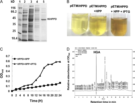
Fig. 7.
(A) SDS–PAGE analysis of over expressed HisTag–MiHPPD fusion protein. Samples were analysed on 10% SDS–PAGE by Coomassie Blue staining. 1, protein marker (MBI-Fermentas); 2, uninduced crude extract; 3, induced crude extract; 4, unbound protein on Ni-NTA column; 5,eluted protein (MiHPPD) using 100 mM imidazol. Position and size of protein markers are shown. (B) Formation of brown pigment in the culture medium of E. coli BL21 (DE3) harbouring pET28a MiHPPD with and with out IPTG. (C) Time-dependent formation of pyromelonin (brown pigment) in cell-free media from oxidation of HGA measured at OD440. (D) Representative HPLC chromatogram showing the synthesis of HGA by E. coli BL21 (DE3) cell extract harbouring the plasmid pET28a MiHPPD. (This figure is available in colour at JXB online.)

