Figure 2.
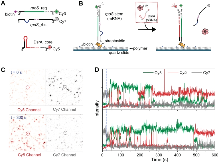
Strand-exchange experiments. (A) RNA fragments used in this study. The three synthetic RNA fragments contain both the RNA–RNA interaction sequences (red line for DsrA_core, and green line for rpoS_reg), and the main Hfq-interaction sequences (DsrA_core: AAUUUUUUAA and rpoS_reg: AUUUUG). The labeling positions of fluorophores and biotin are shown. (B) Experimental scheme. Pre-annealed rpoS_reg:rpoS_rbs complex was immobilized on a polymer-coated surface. While single-molecule fluorescence images were taken, the buffer containing Cy5-labeled DsrA_core (2 nM) and Hfq (5 nM) was delivered. (C) Single-molecule images before and 300 s after the addition of Hfq and DrsA. The circled molecule corresponds to first trace in (D). (D) Single-molecule time traces of Cy3 (green), Cy5 (red) and Cy7 (gray) intensities. Blue dashed line indicates the Hfq injection point.

