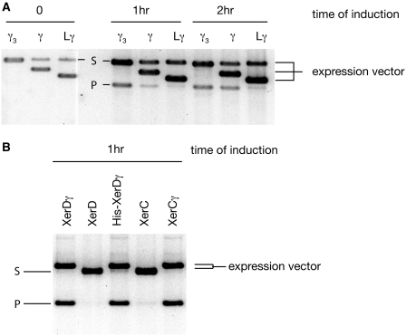
Figure 1.
(A) Timecourse of in vivo recombination of a plasmid with two dif sites in direct repeat. DNA was prepared from cells at the indicated times after induction. Three different constructs expressing variants of isolated FtsKγ domain (γ) cloned into various plasmid backgrounds were used (hence the size differences of the expression vectors). Recombination yields the product P after induction of γ. The smaller, non-replicating product migrates further in the gel and becomes lost from the population over time, so has not been shown. Note that the γ3 expression vector migrates at the same position as the unrecombined reporter plasmid. (B) In vivo recombination following induction of the Xer recombinases and their fusions to γ. DNA was sampled 1 h after induction of each protein. The expression vectors and reporter plasmid migrate at similar positions, but the larger recombination product is clearly visible in the lanes where the fusion proteins are expressed. The stimulation of recombination by the γ fusions to XerC and XerD was so efficient that significant recombination was also observed at the zero time point (not shown) because of leaky expression of the proteins.

