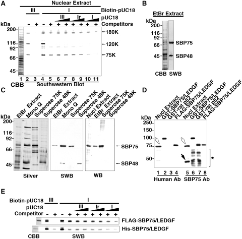
Figure 1.
Supercoiled DNA-binding protein SBP75 is identical to LEDGF/p75. (A) Detection of DNA binding proteins in rat brain nuclear extract by Southwestern blotting. Nuclear extract (75 µl) was subjected to 7.5% SDS–PAGE and separated proteins were blotted on a membrane, which was then incubated with biotin-labeled pUC18 DNA: form III (lanes 2 and 3) or form I (lanes 4–11) in the absence (lanes 2 and 4) or the presence (lanes 3 and 5–11) of non-specific competitors (calf thymus DNA and yeast tRNA). Excess amounts of unlabeled pUC18 DNA: form III (lanes 6 and 7), form Ir (lanes 8 and 9) or form I (lanes 10 and 11) were added at 5-fold (lanes 6, 8 and 10) or 25-fold excess (lanes 7, 9 and 11). Proteins with apparent molecular mass of 180 kDa (180K), 120 kDa (120K) and 75 kDa (75K) are indicated. Total protein on the membrane was stained with CBB (lane 1). (B) Proteins extracted with 20 mM EtBr were subjected to Southwestern blotting (SWB). The blot was incubated with biotin-labeled form I pUC18 in the presence of non-specific competitors. Supercoiled DNA bound to the two proteins labeled SBP75 and SBP48 selectively (right lane). Total protein in the extract was stained with CBB (left lane). (C) Proteins in 20 mM EtBr extract were fractionated by chromatography on Mono Q and Superose columns and pooled fractions were subjected to SDS–PAGE and stained with silver reagent (left panel). The same sets of samples were blotted onto a membrane and analyzed by SWB or western blotting (WB) with human autoantibody to SBP75. (D) Rat nuclear extract and SBP75 recombinant proteins were subjected to western blotting analysis with human autoantibody to SBP75 (Human Ab, lanes 1–4) or with newly prepared anti-rat SBP75/LEDGF polyclonal antibody (SBP75 Ab, lanes 5–8). Open arrows indicate endogenous SBP75 and closed arrow indicates SBP48 that was reactive to polyclonal antibody but not to human autoantibody. Lower bands in GST fusion proteins that were detected by SBP75 Ab (indicated by asterisk in lanes 6 and 7) but not by human Ab are degradation products lacking C-terminal portions. For further analysis of epitope position recognized by human autoantibody, see Supplementary Figure S1. (E) Purified FLAG- or His-tagged SBP75/LEDGF (2.5 µg each) was subjected to Southwestern analysis as in panel A. CBB-stained proteins are shown in the left most lane.

