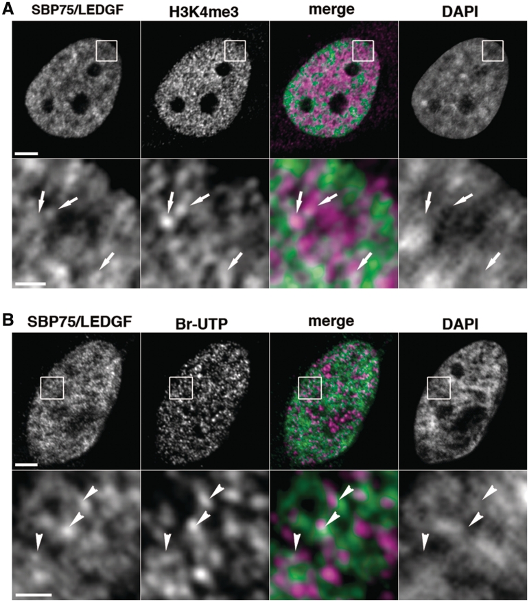
Figure 5.
Intranuclear distribution of endogenous SBP75/LEDGF and active transcription sites in HeLa cells. (A) Cells were fixed with 4% paraformaldehyde and immunostained doubly with affinity purified human autoantibody to SBP75/LEDGF (3 µg/ml) and rabbit polyclonal antibody to human trimethyl-lysin4 in histone H3 (H3K4me3, 1/1000×), followed by incubation with Alexa 488-conjugated goat anti-human IgG (5 µg/ml) and Alexa 594-conjugated goat anti-rabbit IgG (5 µg/ml). DNA was stained with DAPI (0.25 µg/ml). Boxed area in upper panels was magnified and shown in lower panels. Arrows in the area with weak DAPI signal indicate that Alexa 488 (green) and Alexa 594 (magenta) signals are overlapped or juxtaposed in the merged image. Scale bars designate 5 µm (upper panels) and 1 µm (lower panels), respectively. (B) Br-UTP was incorporated into transcription sites in the cells permeabilized with 0.05% Triton X-100 by incubation with other nucleotides and detected with anti-BrdU mouse monoclonal antibody (1 µg/ml) that was visualized with Alexa 594-labeled second antibody. Endogenous SBP75/LEDGF was also detected with affinity purified human autoantibody to SBP75/LEDGF as in panel A. Arrowheads in low DAPI area indicate that Alexa 488 (green) and Alexa 594 (magenta) signals were overlapped or juxtaposed in the merged image. Scale bars designate 2 µm (upper panels) and 0.5 µm (lower panels), respectively.

