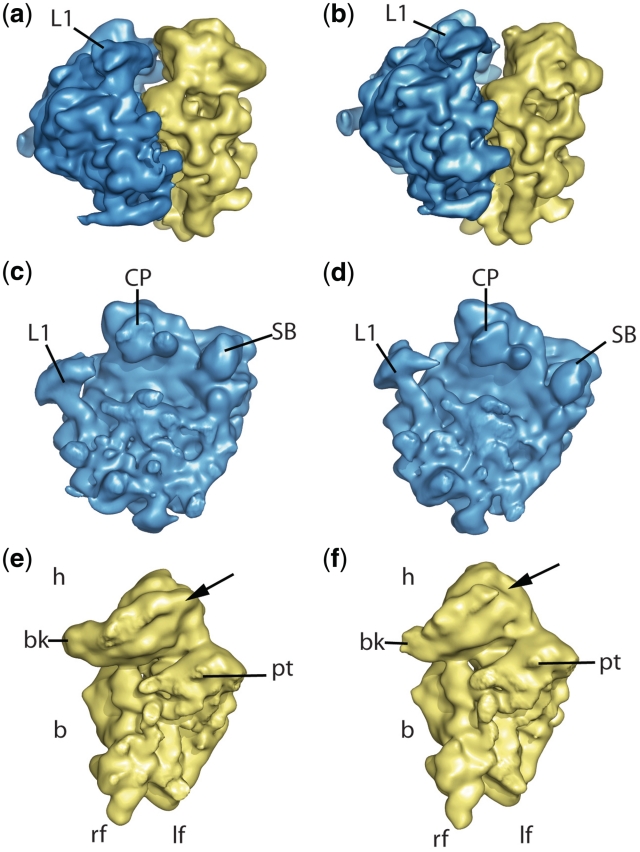
Figure 1.
The cryo-EM maps from the wild-type and rpS25-deletion mutant. (a and b) 80S ribosomes from wild-type and mutant, respectively, seen from the L1 side. (c–f) The corresponding 60S (blue, c and d) and 40S (yellow, e and f) subunits from the interface side. Landmarks of the 40S subunit are head (h), body (b), beak (bk), platform (pt), left and right foot (lf and rf, respectively). Landmarks for the 60S subunit are central protuberance (CP), L1 protuberance and stalk base (SB). The arrow at the 40S subunit points the estimated localization of the protein rpS25.

