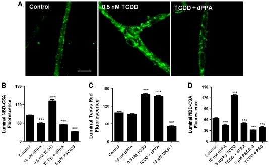
Figure 1.
Activating protein kinase C (PKC)-β1 reverses 2,3,7,8-tetrachlorodibenzo-p-dioxin (TCDD)-induced increases in P-glycoprotein transport activity in rat brain capillaries in vitro and ex vivo. (A) Representative confocal images of control capillaries and capillaries exposed to 0.5 nmol/L TCDD, 0.5 nmol/L TCDD plus 10 nmol/L 2-deoxyphorbol-13-phenylacetate-20-acetate (dPPA) in vitro. Capillaries were incubated for 3 hours in medium without and with TCDD. During the last 30 minutes, some of the capillaries were exposed to dPPA. At the end of the incubation period, (N-∈(4-nitrobenzofurazan-7-yl)-D-Lys8)-cyclosporine A (NBD-CSA; 2 μmol/L) was added to the medium and capillaries were imaged after 1 hour. White scale bar indicates 5 μm. (B) Effects of exposure to TCDD, dPPA, or TCDD plus dPPA on luminal NBD-CSA fluorescence. (C) Effects of exposure to TCDD, dPPA, or TCDD plus dPPA on luminal Texas red fluorescence. Protocol was the same as in A and B, except that Texas red was used to monitor changes in Mrp2 transport activity. (D) Effects of PKC-β1 activation on capillaries isolated from control and TCDD-exposed rats (single intraperitoneal dose, 5 μg/kg; tissue collected after 2 days). Protocol for isolated capillaries is same as in A. Shown are mean±s.e.m. for 8 to 12 capillaries from single preparations, each containing pooled brain tissue from 5 to 10 rats. Statistical comparisons (one-way analysis of variance): significantly different from control, ***P<0.001.

