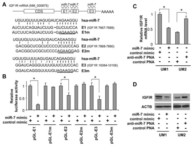
Figure 2. miR-7 direct targeting of IGF1R mRNA.
(A) Schematic representation of IGF1R mRNA showing the positions and sequences of the three predicted miR-7-binding sites located in its 3′-UTR. CDS, coding sequence. (B) Dual-luciferase reporter assays were performed as described in the Experimental section on cells transfected with constructs containing the predicted targeting sequences (pGL-E1, pGL-E2 and pGL-E3) or targeting sequences with mutated seed regions [pGL-E1m, pGL-E2m and pGL-E3m; mutations are underlined in (A)] cloned into the 3′-UTR of the reporter gene, and treated with miR-7 mimic or control mimic. Quantitative real-time RT–PCR (C) and Western blot (D) analyses were performed to examine the effects of miR-7 on IGF1R expression in UM1 cells that were treated with miR-7 mimic or control mimic, and UM2 cells that were treated with anti-miR-7 PNA or scrambled LNA (locked nucleic acid). Results shown in (B) and (C) are means ± S.D.; all results are representative of at least three independent experiments. *P < 0.05.

