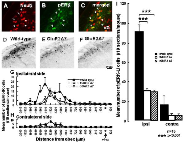
Fig. 2.
(A–C) Photomicrographs of NeuN positive cells (A), pERK-LI cells (B) and pERK-LI cells double stained with NeuN antibody (C) in the wild-type mice. (D–F) Photographs of pERK-LI cells in the wild-type mice (D) and GluR2 7 mutant mice (E) and GluR3 7 mutant mice (F) at 5 min after 2 g mechanical stimulation of the whisker pad skin at 7days after ION partial transaction. The photomicrographs in A–F were taken from sections at 1440 μm caudal to the obex. (G–I) Rostro-caudal distribution (G and H) of pERK-LI cells in the wild-type, GluR2 and GluR3 delta7 KI mice. ((G) Ipsilateral side with ION partial transection; (H) contralateral side with ION partial transection). (I) The mean number of pERK-LI cells in the both sides Vc and C1/C2 ION-transected mice. ***p < 0.001.

