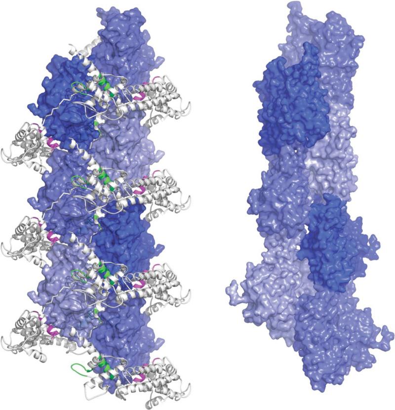
Figure 5.
Precursor helix assembled by the formin FH2 domain. The left view shows the precursor helix assembled by FH2 subunits along the crystallographic C2 axis. The forming FH2 domain is shown grey as in Figure 1i. Neighboring molecules are positioned by binding respectively to the knob (magenta)[ and the post (green) to form a helix with a twist of 180° per molecule and a rise per residue of 28.1 Å (see Supplemental Figure 2 for details). The right view shows the F-actin helix for comparison. Actin subunits of the filament helix are rotated by -166.6° and translated by 27.6 Å (as in Figure 2)

