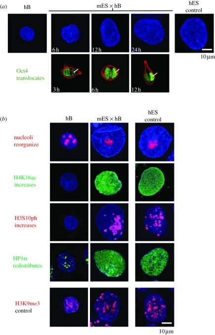
Figure 2.
Remodelling of somatic nuclei during pluripotent reprogramming. (a) DAPI-labelled hB cells before and at sequential times (in hours) after fusion and heterokaryon formation with mouse ES cells (mES × hB) reveal an increased nuclear size (blue) and influx of Oct4 protein (green). Oct4 is detected in the hB (arrowed) nucleus within 3 h (and peaks at 12 h), ahead of human Oct4 transcription [15]. Representative confocal images are shown. (b) Human nuclei before (hB) and 24 h after fusion (mES × hB), labelled to reveal specific nuclear changes that occur early during reprogramming. Human ES cells are shown as the control. Antibodies used detect nucleolar marker B23 (nucleophosmin, red), acetylated H4K16 (H4K16ac, green), phosphorylated H3S10 (H3S10ph, red), HP1α (green) and tri-methylated H3K9 (H3K9me3, red). H3K9me3, which does not appreciably change, is provided as a control. Representative confocal images are shown.

