Figure 3.

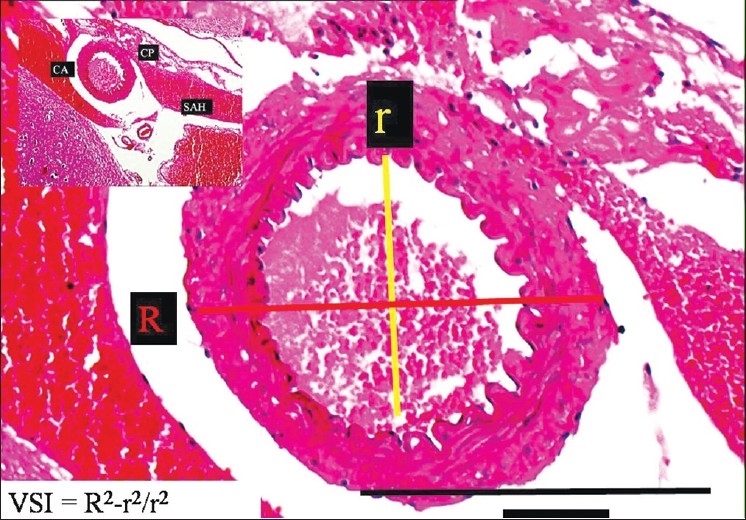
The histopathological appearance of the right AChA at the postorigin level in a SAH rabbit model (LM, H and E, ×100). On the left side, minimal forms of CP are observed (LM, H and E, ×20) (mild vasospasm associated SAH group)

The histopathological appearance of the right AChA at the postorigin level in a SAH rabbit model (LM, H and E, ×100). On the left side, minimal forms of CP are observed (LM, H and E, ×20) (mild vasospasm associated SAH group)