
Figure 3.
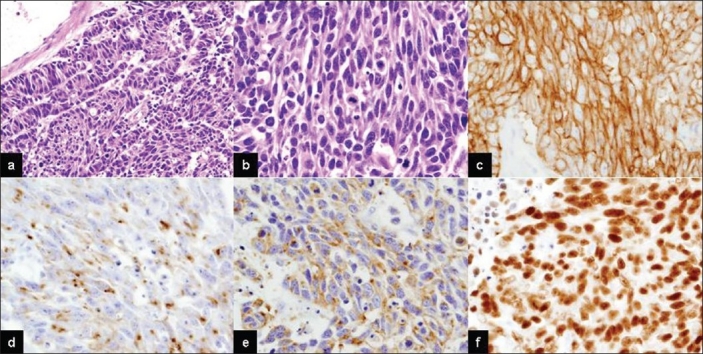
Microscopic findings of metastatic brain large cell neuroendocrine carcinoma. Neoplastic cells showing a sheet-like growth with trabecular and rosette-like patterns (a, b). Tumor cells demonstrating abundant cytoplasm, frequent salt-and-pepper like nuclear chromatin and numerous mitotic figures (H and E: a, ×100; b, ×200). Tumor cell expression of CD56 (c), chromogranin A (d), synaptophysin (e) and TTF-1 (f) (×200)