Figure 2.

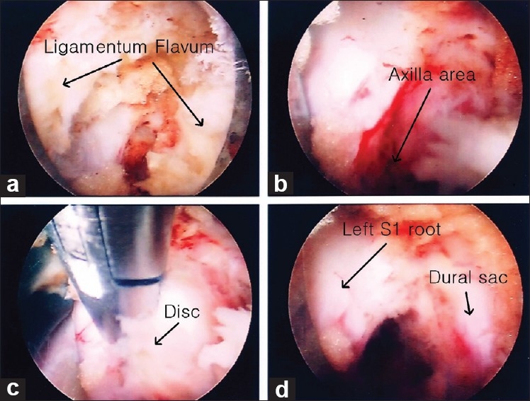
Intraoperative endoscopic view showing the following: (a) opening the ligamentum flavum, (b) exposure of the left axilla area, (c) extruded disc material in the axilla, and (d) dural sac with left S1 root and axilla after decompression

Intraoperative endoscopic view showing the following: (a) opening the ligamentum flavum, (b) exposure of the left axilla area, (c) extruded disc material in the axilla, and (d) dural sac with left S1 root and axilla after decompression