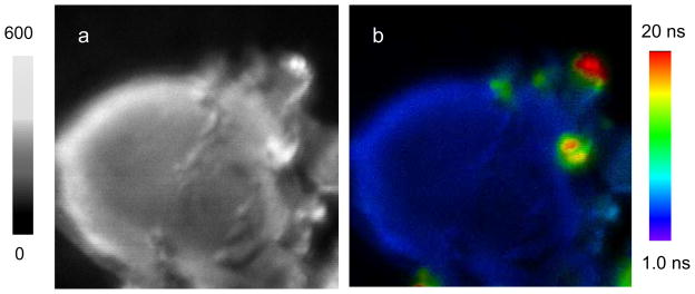
Figure 6.
Representative fluorescence (a) intensity and (b) lifetime cell images conjugated by the Ru(phen-NH2)32+-Ag shells, Ru(bpy)32+-Ag shells, and Ru(dpp)32+-Ag shells upon the excitation at 470 nm. The scales of diagrams are 15 × 15 μm. The resolutions of diagrams are 100 × 100 pixel with an integration of 0.6 ms/pixel.

