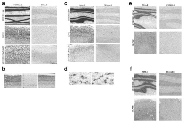
Figure 3.
In situ hybridization of male and female brain sections from the cerebellum, DLPFC, and anterior cingulate cortex. (a) The XIST antisense probe is shown for six sections (three males and three females) with significant pattern of labeling in neurons from female sections. (b) The XIST probe shows female DLPFC hybridization signal (left and right panels) was present in all layers of cortex with laminar-specific distribution and absent in male DLPFC layers (middle panel). In general signal was stronger in superficial layers than in deeper layers for female DLPFC. The laminar distribution and lack of hybridization in white matter suggest that XIST is prevalently expressed in neurons. (c) The 35S-XIST riboprobe silver grains reveals dense labeling of neurons and very sparse and usually absent labeling of glia. (d) The RPS4Y probe shows predominant pattern of labeling in neurons for male tissues. (e) The SMCY probe and UTY probe (f) also confirms the microarray and TaqMan/real-time PCR experiments showing a strong labeling of male cerebellum, which shows higher tissue labeling compared to both cortical regions.

