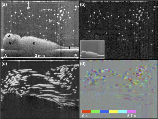
Fig. 4.

OCT imaging of X. tropicalis epithelial cilia-driven flow. (a) B-scan of embryo in microparticle-seeded physiologic solution. Original image stack filtered with a 2x2x2 (x,z,t) pixel filter. (b) Background-subtracted B-mode image. The inset in (b) is the minimum projection image across all B-scans over the 5.7 s acquisition. (c) OCT pathline imaging generated by taking the maximum projection over all background-subtracted images over the 5.7 s acquisition. (d) Color-encoding of time pathline imaging. b, body; e, eye; h, head; m, microsphere; t, tail; w, water-air interface. A, anterior; P, posterior. Scale image to have square pixels. Media 1 (9.3MB, MOV) shows the OCT movie, background-subtracted movie, and related cumulative maximum projection (i.e. cumulative particle pathline) movies.
