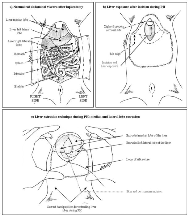
Fig. 24.1.
Partial hepatectomy: (a) Normal anatomy of the rat abdominal viscera. (b) Following incision, three lobes should be clearly visible. (c) The median and left lateral lobes of the liver should be externalized, through the loop of silk suture, by pressure from the thumbs. The index fingers brace the lower portion of the rib cage.

