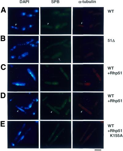
Figure 6.
Indirect immunofluorescence microscopic analysis. Cells overexpressing Rhp51 (C and D) and Rhp51 K155A (E) were collected 25 h after thiamine deprivation and stained with DAPI (left), anti-Sad1 (SPB) (middle) and anti-tubulin (right) antibodies. Wild-type (A) and rhp51Δ (B) cells are also shown. Bar 10 µm.

