Figure 1.
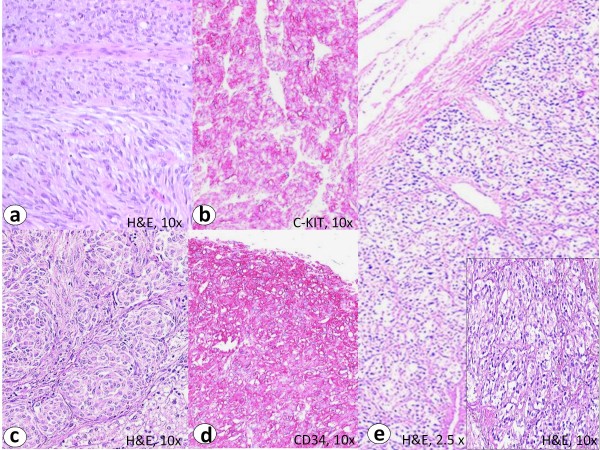
The primary tumor of the CT-GIST (case 1) showed a biphasic histomorphological growth pattern with spindled and epithelioid cells (a, H&E staining). Immunohistochemistry revealed an intensive predominantly membranous staining with C-KIT (b) and CD34 (d). The liver metastases revealed small nests and "Zellballen" of tumor cells with epithelioid growth pattern (c, H&E staining). The paraganglioma showed a characteristic nested pattern (e, H&E staining). Note similarity to hepatic metastasis from GIST in c.

