Figure 2.
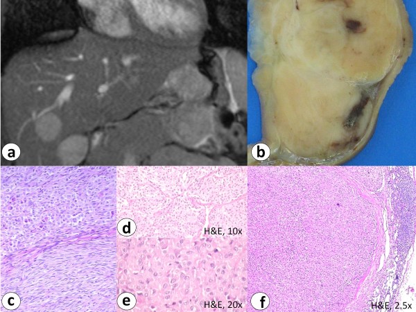
Magnetic resonance tomography in case 2 showed an exophytic, lobulated and ulcerated antral mass diagnosed as gastric GIST with multiple smaller satellite tumor nodules, liver (a, b) and lymph node metastases (f) (H&E staining). Histology reveals biphasic histomorphological growth pattern (c) with fibromuscular septa (d) and hypercellularity (e) (H&E staining).

