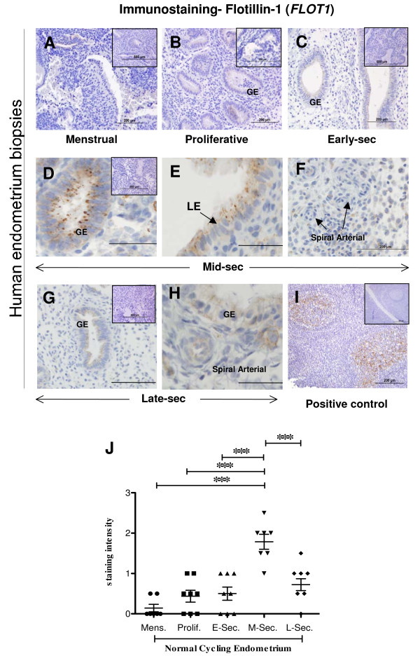
Figure 6.
Photomicrograph representing immunohistochemical staining for flotillin-1 (FLOT1) in human endometrial tissue throughout the menstrual cycle and in ECC-1 cells. Positive staining for FLOT1 showed as brown pigment with blue nuclear counterstain. (A) Menstrual, d1-4 (B) Proliferative, d5-14 (C) Early-secretory, d15-20 (D-F) Mid-secretory, d21-24 (G-H) Late-secretory, d25-d27. (GE) Glandular epithelial, (LE) Luminal epithelial. Insets in each case are matched negative controls. Positive control of human tonsil included (I). Bars represent 200 μm. Semi-quantitative analysis of staining intensity for FLOT1 in GE (J). Data represented as mean ± SEM. * P ≤ 0.05, ** P ≤ 0.01, *** P ≤ 0.001, Kruskal Wallis test, with Dunn's post-hoc test.

