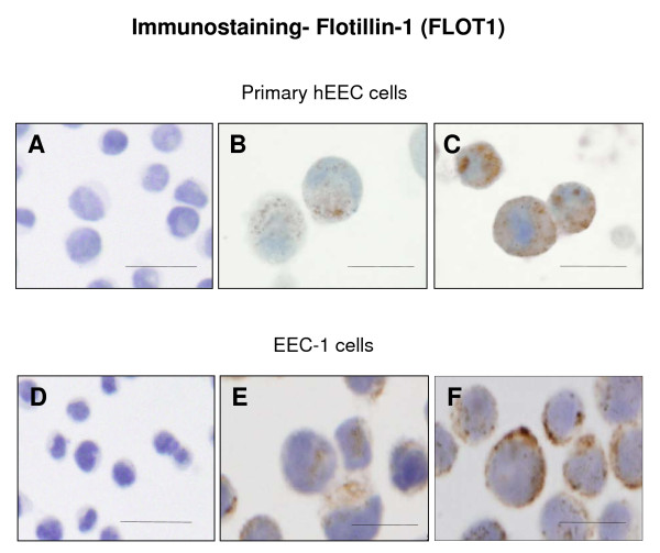
Figure 7.
Photomicrograph representing immunocytochemical staining for flotillin-1 (FLOT1) in human endometrial epithelial cells (hEEC). Primary hEEC and the cell lines ECC-1 were treated with diluent or IL11 (100 ng/ml) for 24 h. Cytospins were prepared and subjected to immunocytochemistry as described in the Materials & Methods. Positive staining for FLOT1 showed as brown pigment with blue nuclear counterstain. (A, D) Negative IgG (B) Control primary EEC (C) IL11 100 ng/ml primary hEEC (E) Control ECC-1 (F) IL11 100 ng/ml ECC-1. Bars represent 200 μm.

