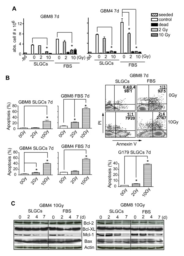
Figure 6.
Delayed γIR-induced apoptosis in SLGCs. SLGCs were irradiated with 0, 2, or 10 Gy. A. 7 d later, the numbers of viable and dead cells were counted after staining with trypan blue. B. Apoptosis was assessed flow cytometrically after staining with Annexin V/PI. A representative flow cytometry analysis is shown (upper right). C. Kinetics of expression of pro- and anti-apoptotic proteins were assessed by Western blotting.

