
Figure 3.
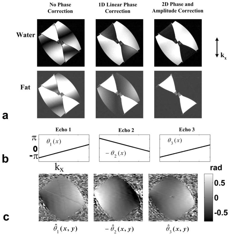
Results from a water–fat phantom scan with obliquely placed slab (approximately 45°) in the coronal plane. a: Separated water and fat images using the three-point bipolar data with no phase correction, 1D linear phase correction, 2D phase and amplitude correction (Nr = 16), respectively (from left to right). b: The estimated linear phase errors in the read-out direction at each echo. c: The high order 2D phase errors at each echo. Due to the anisotropic gradient delays, substantial phase errors appear in the phase encoding direction (horizontal) which cannot be resolved by the 1D phase correction.