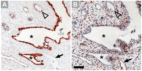
Figure 3. Comparison of localization of IDO1 (A) and HLA-DR (B) in first trimester decidua.
Serial paraffin sections are stained by immunohistochemistry. Designated are uterine glands (asterisks), the endothelium of spiral arteries (open arrowhead) and of veins (arrow). Please note that apart from expression in the endothelium of veins, HLA-DR is expressed by macrophages. Scale bar = 100 µm.

