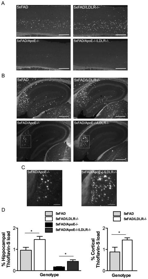
Figure 1. LDLR deficiency results in increased Thioflavine-S plaque load in the presence or in the absence of ApoE.
A–B. Representative pictures of Thioflavine-S staining in cortices (A) and hippocampi (B) of the analyzed groups (n = 5−7, 6–7 sections per animal, 240 mm apart). The absence of LDLR results in increased Thioflavine-S positive staining in the 5XFAD mice in the presence (A and B upper photos) or the absence of ApoE (A and B lower photos). Scale bar 0.5 mm. C. Magnification of the subiculum of the 5XFAD/ApoE-/- and the 5XFAD/ApoE-/-LDLR-/- mice, showing the difference in abundance of Thioflavine-S positive plaques. Scale bar 0.1 mm. D. Quantitation of Thioflavine-S positive staining in the hippocampi (left) and cortices (right) of female mice showing the increase in the amyloid plaques in the 5XFAD/LDL-/- and 5XFAD/ApoE-/-LDLR-/- mice. One-way ANOVA showed a significant difference among groups (P<0.0001) followed by Student's t-test. * P<0.05. P-values among groups are analysed in Table 1.

