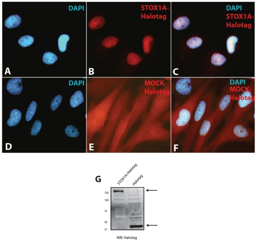
Figure 6. Expression analysis of STOX1A in stable transfected U-373 MG cells.
(A, –C) Immunofluorescence shows primarily nuclear staining for STOX1A-Halotag protein in stable transfected U-373 MG cells while stable MOCK-Halotag transfected U-373 MG cells shows diffuse cytoplasmatic staining (D, –F). (G) Expression of STOX1A protein was determined with an anti-Halotag specific antibody by western blot using total cell protein extracts obtained from stable STOX1A (left lane) and MOCK (right lane) transfected U373 cells. A specific band representing STOX1A-halotag protein was observed at its expected size of 150 kd. Halotag (MOCK) protein was detected at its expected size of 34 kd. Westernblot image is a representative of at least 3 independent experiments.

