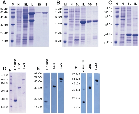
Figure 3. Protein analysis by SDS-PAGE and Western blotting.
(A) Lsa66, (B) Lp30 and (C) rLIC12238 expression from NaCl-induced E. coli Bl21-SI. M: molecular mass protein marker; NI: non-induced total bacterial extract; SL: supernatant after bacterial cell lysis and centrifugation; IL: inclusion body pellet after bacterial lysis and centrifugation; SS: soluble fraction of the induced culture in the presence of 8 M urea; IS: insoluble fraction of the induced culture in the presence of 8 M urea. (D) Comassie blue stained purified recombinant proteins. (E) and (F) are western blotting analysis of the recombinant proteins probed with anti-His tag antibodies and the respective homolog antiserum, respectively.

