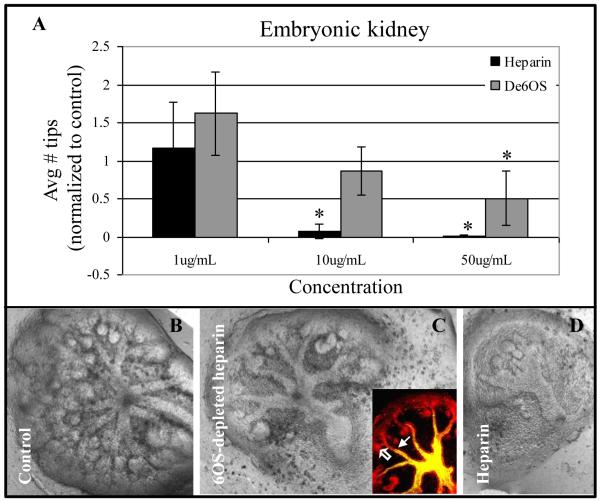
Figure 2.
Effect of 6OS-depleted heparin on embryonic kidney branching morphogenesis. A: Graphical analysis of the average number of UB tips as a percentage of control when cultured in the presence of varying concentrations of heparin or 6OS-depleted heparin. Mean ± SD, n>3, *p<0.05 compared to control. B-D: Phase contrast photomicrographs of whole embryonic kidneys cultured for 4-6 days in the absence (B) or presence of 100 μg/ml of either de6OS-heparin (C) or heparin (D). Even at higher concentrations, compared to heparin-treated kidneys (C), UB architecture is largely preserved despite further reduction in UB tip number in the presence of de6OS (B; inset is a confocal image of the embryonic kidney; open arrow points to UB tip and closed arrow points to UB stalk (D. biflorus lectin (green) and E-cadherin (red)). Photomicrographs taken at 10X magnification.

