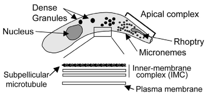
Fig. 2.

Typical zoite cellular organization. Cryptosporidium zoites share a similar body plan to other Apicomplexans including: a crescent shaped cell body; apically localized rhoptry and micronemes; and numerous dense granules throughout. The parasite pellicle consists of an outer plasma membrane and an inner membrane complex composed of 2 distinct membranes with an underlying array of subpellicular microtubules. Zoite gliding motility requires the secretion of adhesive molecules from the apical pole of the parasites (e.g. TRAP) that adhere to substrate receptors; posterior translocation of the adhesive molecules in an actomyosin-dependent manner; and iii) proteolytic cleavage and release of the parasite molecule in motility trails. Zoite stylized from [58].
