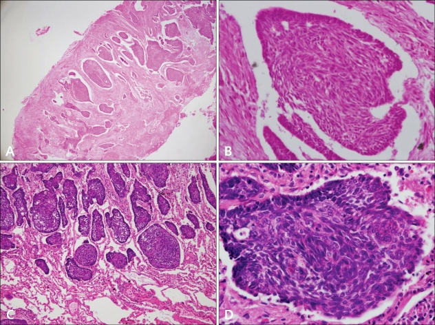
Fig. 3.
(A) Pathological examination of the excised pulmonary nodule demonstrated irregularly shaped tumor masses and retraction of the stroma around the tumor islands (H&E, ×40). (B) The tumor from the lung was composed of basaloid cell nests with peripheral palisading (H&E, ×400). (C) The patient's left maxilla BCC excised 10 years earlier showed similar histopathology with pulmonary MBCC (H&E, ×40). (D) The tumor cells from left maxilla are basaloid cells showing peripheral palisading (H&E, ×400).

