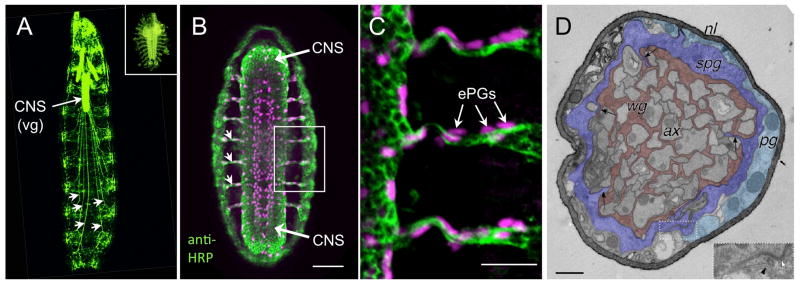
Figure 1.
Anatomy of the larval nervous system. A. An intact 2nd instar larva expressing GFP in the nervous system. The abdominal nerves (some of which are marked with arrowheads) are hundreds of microns long, linking the ventral ganglion (vg) to the periphery. Inset: Nervous system of an embryo (fillet prep) to the same scale as the larva, stained with the neuronal marker anti-HRP. B. Whole embryo stained with anti-HRP (green) and anti-repo (magenta). At this stage the precursors of the subperineurial glia, the embryonic peripheral glia (ePG, magenta) are migrating along the nerves (green), which are less than 20 μm long. Bar: 50 microns. C. Enlarged view of the boxed area in B shows the close association of the ePG (some of which are indicated by arrows) with the nerves Bar: 20 microns. D. TEM of a 3rd instar larval abdominal nerve, showing the four cellular components: axons (ax), wrapping glia (wg), subperineurial glia (spg), and perineurial glia (pg). The inset shows an example of an autocellular septate junction between two processes of a SPG (with permission from Stork et al. 2008). Bar: 1 micron.

