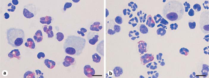
Fig. 4.
Representative photomicrographs of BALF cells in animals given i.n. HDM sensitization and challenge (Diff-Quick staining). The percentage of eosinophils was higher in TRPV1 receptor gene knockout mice (a) than in wild-type animals (b). Scale bars = 10 μm. The eosinophils are indicated by red staining.

