Fig. 5.
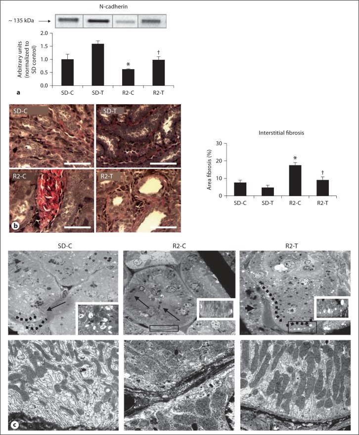
Angiotensin II contributes to loss of PT N-cadherin, basolateral remodeling and tubulointerstitial fibrosis. a Western blot analysis ofthe PT-specific adhesion molecule N-cadherin. b Verhoeff-Van Gieson (VVG) stain for elastin and collagen with measures of tubulointerstitial fibrosis to the right. Scale bar 50 μm. * p < 0.05 when compared to age-matched Sprague-Dawley controls (SD-C); † p < 0.05 when telmisartan-treated R2 rats (R2-T) are compared to age-matched Ren2 controls (R2-C). c Representative images from ultrastructural analysis of TEM for the S-1 region of the PT. Top panel depicts lysosomes in the basal region of the PT wherein there is loss of electron dense lysosome (arrows) in the R2-C model (middle panel) compared to age-matched SD-C, restored with telmisartan treatment in the Ren2. Bottom panel depicts elongated canalicular plasma membrane infoldings, loss of basal polarity, elongated mitochondriaand basement membrane thickening in the basal region of the R2-C compared to SD-C, findings improved in the telmisartan-treated R2 model.

