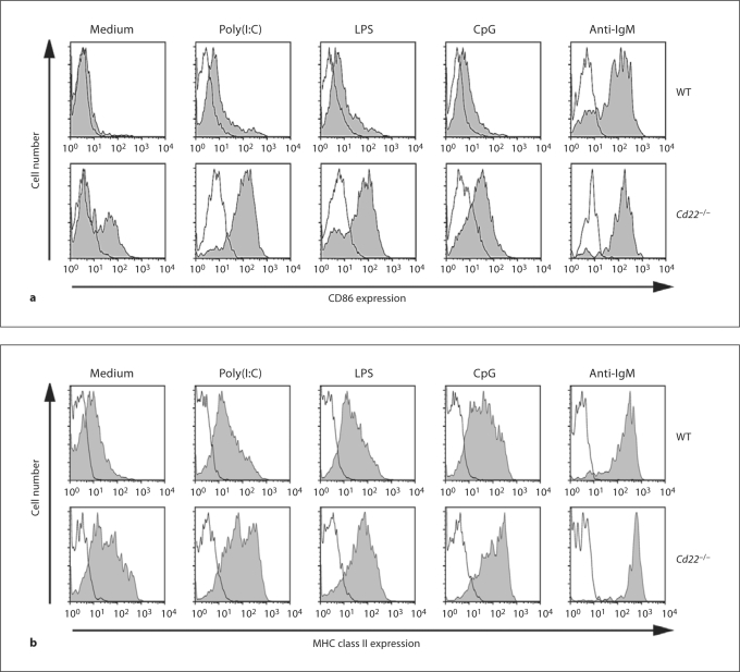
Fig. 2.
Augmented CD86 and class II induction in Cd22–/– B cells in response to TLR ligands. WT and Cd22–/– B cells were stimulated for 2 days with 10 μg/ml poly(I:C), 0.15 μg/ml LPS, 7.5 n M CpG and 0.3 μg/ml anti-IgM. Then cells were harvested and stained with anti-CD86 (a) or anti-MHC class II I-A b (b) (gray) or isotype-matched control Ab (black). Stained cells were washed and analyzed by flow cytometry. Data are representative of 3 independent experiments with similar results.

