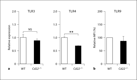
Fig. 3.
TLR expression in Cd22–/– B cells is not significantly different from that of WT B cells. a Total RNA was extracted from isolated WT and Cd22–/– B cells. Expression of TLR3 and 4 was examined by quantitative real-time PCR. Expression levels were normalized to β-actin. Statistical analyses were performed by Student's t test. b Isolated B cells were fixed, permeabilized and then stained with anti-TLR9 Ab followed by R-phycoerythrin-labeled secondary Ab. Stained cells were washed and analyzed by flow cytometry. Geometric mean fluorescence intensity (MFI) of Cd22–/– B cells relative to WT is shown. Statistical analyses were performed by Student's t test. Data are representative or means of 3 independent experiments with similar results.

