Figure 2.

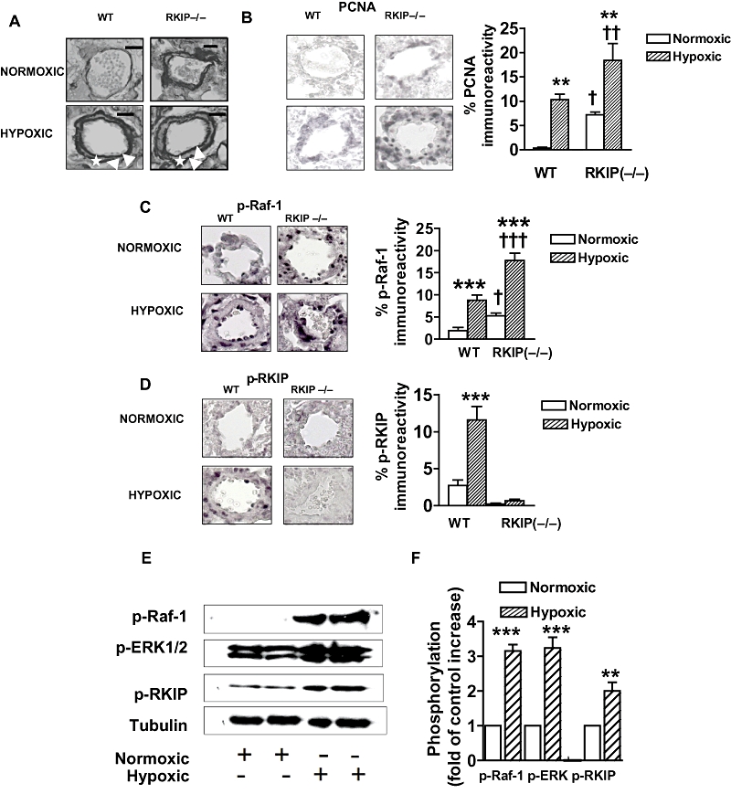
Representative photomicrographs showing the effects of chronic hypoxia and deletion of the Raf-1 kinase inhibitor protein (RKIP) gene (A–D) on: (A) pulmonary vascular remodelling and (B) expression of proliferating cell nuclear antigen (PCNA), (C) expression of phospho-Raf-1, (D) phospho-RKIP in small pulmonary arteries from mice and the effect of chronic hypoxia on (E) and (F) the RKIP signalling elements in small pulmonary arteries from wild-type (WT) mice. (A) The figure demonstrates that remodelling occurs in the normoxic RKIP (−/−) group and is exaggerated in hypoxia when compared with WT mice. Note the readily identifiable double elastic lamina (white arrow) and newly formed smooth muscle layer (asterisk) in the hypoxic groups. (B) The figure demonstrates that expression of PNCA is increased in the pulmonary arteries of the mice lacking RKIP [RKIP (−/−)], indicative of increased proliferation. Hypoxia-induced proliferation is also observed in the WT mice which is further exaggerated by hypoxia in the RKIP (−/−) mice. (C) The figure demonstrates hypoxia-induced phosphorylation of Raf-1 (p-Raf-1; shown as black staining) in the WT mice which is further exaggerated by hypoxia in the RKIP (−/−) mice. D. The figure demonstrates hypoxia-induced phosphorylation of RKIP [shown as black staining; absent in the RKIP (−/−) mice] in the WT mice. (E,F) The figure demonstrates that chronic hypoxia induces phosphorylation of the RKIP signalling elements Raf-1, RKIP and ERK1/2. Scale bars = 25 µm. *P < 0.05, **P < 0.01, ***P < 0.001 significantly greater than corresponding value in normoxic mice. †P < 0.01, ††P < 0.001, significantly greater than corresponding value in WT mice. Data are shown as mean ± SEM.
