
Figure 1.
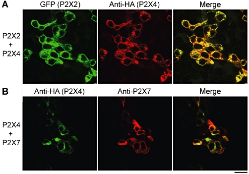
Co-expression of P2X receptors in tsA 201 cells. (A) Cells were co-transfected with DNA encoding P2X2-GFP and P2X4-HA. Cells were fixed, permeabilized and incubated with a mouse monoclonal anti-HA antibody, followed by Cy3-conjugated goat anti-mouse secondary antibody. Cells were imaged by confocal laser scanning microscopy. The left-hand panel shows the GFP signal, the centre panel the Cy3 signal and the right-hand panel the merged signals. Cells expressing GFP and Cy3 completely coincide, indicating that all of the transfected cells express both P2X2 and P2X4. (B) Cells were co-transfected with DNA encoding P2X4-HA and His10-P2X7. Cells were fixed, permeabilized and incubated with mouse monoclonal anti-HA and rabbit polyclonal anti-P2X7 antibodies, followed by either FITC-conjugated goat anti-mouse or Cy3-conjugated goat anti-rabbit secondary antibodies. The left-hand panel shows the FITC signal, the centre panel the Cy3 signal, and the right-hand panel the merged signals. Cells expressing FITC and Cy3 extensively coincide, indicating that the vast majority of transfected cells express both P2X4 and P2X7 receptors. Scale bar, 25 µm. FITC, fluorescein isothiocyanate; GFP, green fluorescent protein; HA, haemagglutinin.