
Figure 3.
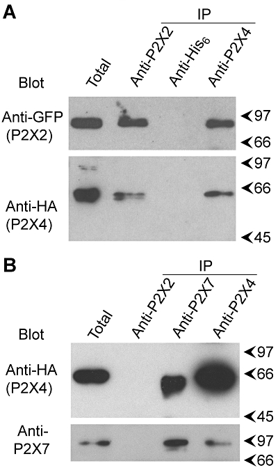
Co-immunoprecipitation of P2X subunits. (A) P2X2-GFP and P2X4-HA were co-expressed by transient transfection of tsA 201 cells. Crude membrane fractions prepared from the cells were solubilized in 1% CHAPS. P2X2-GFP was immunoprecipitated using a rabbit polyclonal anti-P2X2 antibody, and P2X4-HA was immunoprecipitated using a rabbit polyclonal anti-P2X4 antibody. A rabbit polyclonal anti-His6 antibody was used as a negative control. Immunoprecipitates were analysed by SDS-PAGE followed by immunoblotting with either mouse monoclonal anti-GFP (P2X2-GFP) or anti-HA (P2X4-HA) antibodies. Immunoreactive bands were visualized using enhanced chemiluminescence. Arrowheads indicate molecular mass markers (kDa). (B) P2X4-HA and His10-P2X7 were co-expressed by transient transfection of tsA 201 cells. P2X4-HA was immunoprecipitated from a detergent-solubilized membrane fraction using a rabbit polyclonal anti-P2X4 antibody, and His10-P2X7 was immunoprecipitated using a rabbit polyclonal anti-P2X7 antibody. A rabbit polyclonal anti-P2X2 antibody was used as a negative control. Immunoprecipitates were analysed by SDS-PAGE followed by immunoblotting with either a mouse monoclonal anti-HA antibody (P2X4-HA) or a rabbit polyclonal anti-P2X7 antibody. CHAPS, 3-[(3-cholamidopropyl)dimethylammonio]-1-propanesulphonate; GFP, green fluorescent protein; HA, haemagglutinin; PAGE, polyacrylamide gel electrophoresis.