
Figure 4.
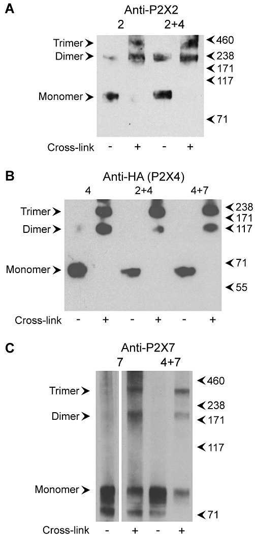
Cross-linking analysis of P2X subunits. (A) Detergent extracts of membrane fractions from cells transfected with either P2X2-GFP alone or P2X2-GFP+P2X4-HA were incubated with the chemical cross-linker DSS (0.2 mM, 30 min at room temperature), or with the DMSO vehicle alone, and then subjected to SDS-PAGE followed by immunoblotting with a rabbit polyclonal anti-P2X2 antibody. Immunoreactive bands were visualized using enhanced chemiluminescence. Arrowheads indicate molecular mass markers (kDa). (B) Detergent extracts of membrane fractions from cells transfected with either P2X4-HA alone, P2X2-GFP+P2X4-HA or P2X4-HA+His10-P2X7 were incubated with DSS (4 mM) and then subjected to SDS-PAGE followed by immunoblotting with a mouse monoclonal anti-HA (P2X4-HA) antibody. (C) Detergent extracts of membrane fractions from cells transfected with either His10-P2X7 alone or P2X4-HA+His10-P2X7 were incubated with DSS (0.2 mM) and then subjected to SDS-PAGE followed by immunoblotting with a rabbit polyclonal anti-P2X7 antibody. DSS, disuccinimidyl suberate; GFP, green fluorescent protein; HA, haemagglutinin; PAGE, polyacrylamide gel electrophoresis.