
Figure 3.
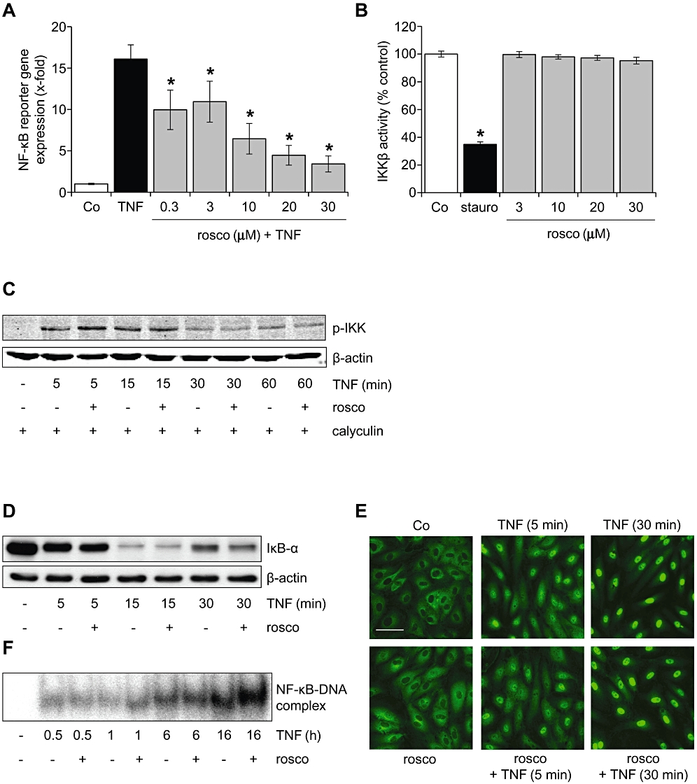
Roscovitine blocked NF-κB-dependent gene expression, but did not influence the NF-κB activation cascade. (A) HUVECs were either left untreated (Co) or pretreated with roscovitine (30 min) and subsequently treated with TNF-α (10 ng·mL−1) for 6 h. Levels of NF-κB-dependent gene expression were analysed by dual luciferase reporter gene expression. n = 3. *P < 0.05 versus TNF-α. (B) The influence of roscovitine on the activity of recombinant IKKβ was assessed by an in vitro kinase assay. Staurosporine (stauro, 100 µM) served as positive control. n = 3. *P < 0.05 versus Co. (C–F) HUVECs were either left untreated (Co) or pretreated with roscovitine (30 min, 10 µM) and subsequently treated with TNF-α (10 ng·mL−1). One representative image out of three independently performed experiments is shown for each assay. (C) Levels of phospho-IKKα/β and β-actin were assessed by Western blot analysis. HUVECs were additionally treated with the protein phosphatase inhibitor calyculin (1 nM) to stabilize the phosphorylation. (D) Levels of IκB-α and β-actin were assessed by Western blot analysis. (E) The NF-κB p65 subunit was visualized by immunocytochemistry and confocal microscopy. White bar = 20 µm. (F) NF-κB DNA-binding activity was determined by electrophoretic mobility shift assay (EMSA). HUVECs, human umbilical vein endothelial cells; IKK, IκB kinase; NF-κB, nuclear factor-κB; TNF-α, tumour necrosis factor-α.