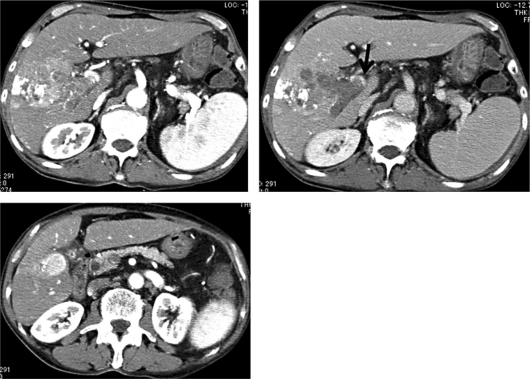
Fig. 3.
Dynamic CT findings of tumor progression. In the main tumor, lipiodol accumulation was observed in some areas; however, tumor enhancement indicated progression in size (top left). The PVTT invaded into the main stem of the portal vein (top right; arrow). The tumor in segment 5 also increased in size (from 2 to 2.4 cm).

