Figure 6.
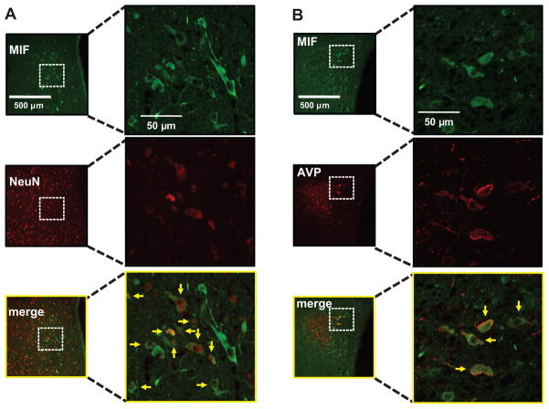
AAV2-CBA-MIF–induced expression of MIF in AVP neurons in the PVN. Wistar rats were injected bilaterally into the PVN with AAV2-CBA-MIF, as detailed in the Methods section. A, Micrographs of MIF and NeuN immunostaining from the same field of the PVN 10 days after microinjection of AAV2-CBA-MIF. Merge: MIF+NeuN. Yellow arrows indicate coexpression of MIF and NeuN. B, Representative fluorescence micrographs of showing MIF and AVP immunostaining from the same field of the PVN 10 days after microinjection of AAV2-CBA-MIF. Merge: MIF+AVP. Yellow arrows indicate coexpression of MIF and AVP.

