Figure 4.
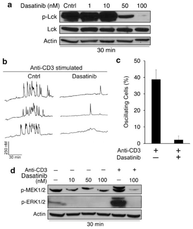
Dasatinib inhibits Lck phosphorylation and TCR signaling. (a) WEHI7.2 cells were treated with vehicle (0.01% DMSO) or dasatinib for 30 min. Phosphorylated (Y394) and total Lck levels were measured by western blotting. β-Actin was used as a loading control. (b) Single cell calcium measurements in WEHI7.2 cells treated with 100 nM dasatinib for 30 min and stimulated with anti-CD3 antibody (2 μg/ml) 1 min into the calcium trace. Representative traces are shown. (c) Quantification of oscillating cells (≥ three spikes) in b. (d) Cells were treated with dasatinib as in a, and phospho-MEK and ERK levels were measured by western blotting. Cells were stimulated with anti-CD3 (2 μg/ml) for 5 min to induce MEK and ERK phosphorylation. Data are representative of multiple independent experiments. Error bars represent the S.E.M.

