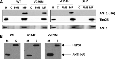
Figure 2.
Localization of human ANT1 in the inner mitochondrial membrane. (A) C2C12 myotubes (DM4) expressing ANT1 (WT, A114P and V289M) or GFP were harvested 48 h after transduction to obtain enriched mitochondrial and cytosolic fractions (C). Mitochondria were further subfractionated into mitoplasts (MP), containing IMs and matrix material, and post-mitoplast supernatant (PMS), containing IMS material and outer membranes. Equal amount of proteins from each fraction were resolved by SDS-PAGE, and immunoblotted for exogenous ANT1 with an antibody against HA (top panel). Tim23 was used as a loading control of inner mitochondrial membrane proteins. The membranes were stripped and re-probed with an ANT1 antibody to determine total levels of ANT1. Purified mitochondria from mouse heart (H) were loaded as a positive control. (B) Mitochondrial fractions were treated with alkaline solution to separate membrane-bound (M) and soluble proteins (S). ANT1-HA was exclusively found in the membrane fraction, whereas the soluble matrix protein Hsp60 was completely released in the supernatant.

